Sunday, October 22, 2023

Stem Cells Treating Rheumatoid Arthritis


As someone who's family has dealt with having a loved one deal with rheumatoid arthritis, I have seen them run through a wide array of treatment options. They tried everything from chemotherapeutic injections to dietary changes and natural remedies, however the only treatment that has worked so far is stem cell treatment. According to an article published by Frontiers in Immunology journal, Mesenchymal stem cell (MSC) treatment uses embryonic mesodermal stem cells for their regenerative properties to promote healing in the recipient. The MSCs secrete a regenerative bioactive factor that acts as a signal to surrounding cells. These factors stimulate site-specific and tissue-specific stem cells of the patient rather than growing into tissue producing cells. Thus, initiating growth of local cells native to the patients own body.

            MSC’s are found naturally in a few main areas of the body which include bone marrow, the umbilical cord, adipose tissue and synovial membranes (the layer surrounding joints which can be affected by rheumatoid arthritis and osteoarthritis). Each of these areas where MSCs are found have distinct advantages and disadvantages in treatment applications. Bone marrow derived MSCs (BM-MSCs) was the first site used for extraction in research studies due to its safety and effectiveness. BM-MSCs are marked by their high differentiation potential which is associated with a high repair potential. However, the disadvantages of BM-MSC’s include the risk of harvesting, condition of the donor, and risk of infection. In the case of the use of these stem cells to treat rheumatoid arthritis, the goal is to reduce the synovial inflammation which leads to synovial hypertrophy, ultimately resulting in bone and cartilage erosion. All these processes leave the patient with severe joint pain and a lack of mobility. In a non-arthritic joint T-lymphocytes and B cells monitor and control inflammation and swelling. When treated with BM-MSCs, however, studies have shown promising progress in the effectiveness of inflammation reduction. Additionally, studies have found that MSCs might be even more effective in treating the swelling of synovial fluid by promoting stimulation of regulating immune cells. Ultimately having a positive outlook on treatment options for subsiding swelling in joints and preventing further degradation of bone and cartilage tissues.

Hwang JJ, Rim YA, Nam Y, Ju JH. Recent Developments in Clinical Applications of Mesenchymal Stem Cells in the Treatment of Rheumatoid Arthritis and Osteoarthritis. Front Immunol. 2021 Mar 8;12:631291. doi: 10.3389/fimmu.2021.631291. PMID: 33763076; PMCID: PMC7982594.

Wednesday, October 18, 2023

Animal Preclinical Models Show Evidence behind "Chemo-Brain"

    Chemo-Brain is the colloquial term for a side effect of chemotherapy treatment that is only commonly talked about amongst cancer survivors. It is the impairment of memory, attention, and cognitive processing that occurs once someone has been exposed to a chemotherapeutic agent. Chemo-Brain is a condition that can be related to by most all cancer survivors yet there is not much research that has been conducted on it. Many survivors have been told that they are not actually experiencing it while others have had to quit jobs or change lifestyles in order to accommodate for it. Any ongoing and recent research about Chemo-Brain needs to be given attention so that cancer survivors and chemotherapy patients with these symptoms can feel validated. One of the strongest pieces of evidence behind Chemo-Brain is a research article published in 2021 called "Animal models of chemotherapy-induced cognitive decline in preclinical drug development". Researchers John et al were able to find evidence in animal models regarding a correlation between chemotherapy exposure and cognitive decline. They found that there were altered neurotransmitter levels, microtubule disintegration, neuroinflammation, reduced neurogenesis, and mitochondrial dysfunction that were found in each of the animals that were exposed to chemotherapeutic agents regardless of which agent was used. This is all to say that the brains of the animals became inflamed, were not able to form new connections between neurons as efficiently and were overall reduced the function of the brain. This all occurred because the chemotherapeutic drugs used caused higher amounts of cytokines in the brain. These cytokines can cause inflammation, so the brain cells became too compacted together and were therefore not able to function at full capacity (John et al., 2021). This research must be brought into human trials to see if this occurs in human patients undergoing chemotherapy.


References

John, J., Kinra, M., Mudgal, J., Viswanatha, G. L., & Nandakumar, K. (2021). Animal models of chemotherapy-induced cognitive decline in preclinical drug development. Psychopharmacology, 238(11), 3025–3053.

Using Adderall Responsibly

    It is not uncommon for people in today's fast-paced world to seek ways to improve their focus, concentration, and overall cognitive performance with Adderall. Prescription drugs containing amphetamine and dextroamphetamine are marketed as Adderall, and they influence the brain's neurotransmitters by stimulating the central nervous system. This drug is mainly prescribed to treat ADHD, which helps patients with these conditions become more focused and control their impulsivity. Due to its Schedule II status as a controlled substance, Adderall carries a risk of abuse and dependence. Once a proper diagnosis has been established, it must be used under the close supervision of a healthcare provider. For people with ADHD, when taken as directed, it can have a significant positive impact and help them live more focused and productive lives. The misuse of Adderall is among the major issues surrounding the drug. To enhance their cognitive abilities or maintain alertness and wakefulness for prolonged periods of time, some individuals use the medication over-the-counter. Misuse of prescription stimulants can result in serious health problems like heart attacks, seizures, elevated blood pressure, and even lethal overdoses. It also discusses the possibility of psychological dependence, since some people may grow very dependent on the drug in order to stay focused or get high. Even when used as directed, Adderall can have negative effects, just like any other drug. These adverse effects may include: nervousness or anxiety; insomnia; elevated blood pressure and heart rate; decreased appetite; digestive problems; and the possibility of mood swings. It is imperative that patients follow the recommended dosage and usage guidelines to reduce the likelihood of Adderall misuse. Through raising awareness of the potential risks of abuse, public awareness campaigns and educational initiatives can encourage the responsible use of Adderall. Adderall is a helpful medication for those who genuinely need it to treat conditions like narcolepsy and ADHD. When used as prescribed, it can lead to significant improvements in focus and overall quality of life. Abuse of Adderall, however, can become addictive and carries a significant risk to one's health. Remember that appropriate usage, precise diagnosis, and consistent contact with medical professionals are crucial for safely optimizing this medication's benefits while lowering its risks. Understanding the truth about Adderall is ultimately the first step to safeguarding your health and well-being if you are considering using the medication.

NIDA. 2018, June 6. Prescription Stimulants DrugFacts. Retrieved from https://nida.nih.gov/publications/drugfacts/prescription-stimulants on 2023, October 18

Mindful-based Interventions within the Workplace

The topic that I was interested in was looking at stress and some ways to manage it. Especially within the workplace, but I believe that a technique like this should be applied to a lot of different places where stress levels are high like schools. Mindfulness-based interventions (MBI) is a time of thoughtfulness that comes from the Buddhist idea of mindfulness. This entails for the person to aware of their thoughts, feelings, bodily sensations and surrounding environment. Never really attaching yourself to any ideas and just being in tune with your surroundings. It's related to being accepting, compassionate, and nonjudgmental. This is done by controlling your breathing, walking, or sitting, and really focusing on filtering out your thoughts. The tradition of mindfulness is becoming something that is not just a Buddhist tradition, but a popular technique that is being used in the educational setting and healthcare. In this research, they looked at the cumulation of all the publications about MBIs and their effects on stress disorders like depression, anxiety, insomnia, eating disorders, addiction, ADHD, and PTSD. They also looked at chronic diseases that affect physical health like pain, hypertension and cardiovascular disease, diabetes, cancer and asthma.

The implication of MBIs is heavily discussed in ethics and the research points out the use of this in business and military settings which may be a cause for concern due to the morals that are encouraged through this technique. However, in a general context, MBI was shown to have a great benefit in helping common mental, physical and social health conditions. I believe that a system like MBI should be implemented in high stress jobs and settings in order to create better workplace environment and happier contributors to society. Happier people means better work efficacy which means more work being done. 


Sources:

   Rachael A. Heckenberg a, a, b, AbstractObjectiveMindfulness-based interventions (MBI) have been related with improved job satisfaction, Kivimäki, M., Niedhammer, I., Eddy, P., Eller, N. H., Bakker, A. B., Grossman, P., Irving, J. A., Lundberg, U., Adam, E. K., Thayer, J. F., Brown, K. W., Jadad, A. R., Kabat-Zinn, J., Malarkey, W. B., Gex-Fabry, M., … O’Leary, K. (2018, September 22). Do workplace-based mindfulness meditation programs improve physiological indices of stress? A systematic review and meta-analysis. Journal of Psychosomatic Research. https://www.sciencedirect.com/science/article/abs/pii/S0022399918305749?via%3Dihub

    Zhang, D., Lee, E. K. P., Mak, E. C. W., Ho, C. Y., & Wong, S. Y. S. (2021). Mindfulness-based interventions: an overall review. British medical bulletin138(1), 41–57. https://doi.org/10.1093/bmb/ldab005

Narcolepsy

One interesting physiological issue is narcolepsy, a rare and intriguing neurological disorder characterized by excessive daytime sleepiness, sudden and uncontrollable episodes of falling asleep (known as cataplexy), and disruptions in the sleep-wake cycle. Narcolepsy is thought to result from a deficiency in the neurotransmitter hypocretin (or orexin), which plays a crucial role in regulating wakefulness and sleep. This condition can significantly impact a person's daily life, making it challenging to stay awake and alert during the day, leading to accidents and difficulties with work or social activities. 


Narcolepsy is estimated to affect about 1 in 2,000 people, and its exact cause remains a subject of ongoing research. The most common form of narcolepsy, Type 1 narcolepsy, is often associated with the loss of hypocretin-producing cells in the brain. Various studies have contributed to our understanding of this condition, such as "Narcolepsy and Low CSF Hypocretin (Orexin) Concentration in Monozygotic Twins" published in the New England Journal of Medicine in 2000. Further research is essential to develop more effective treatments and management strategies for narcolepsy, ultimately improving the quality of life for individuals living with this condition. 


Source: Chemelli, R. M., Willie, J. T., Sinton, C. M., Elmquist, J. K., Scammell, T., Lee, C., ... & Yanagisawa, M. (1999). Narcolepsy in orexin knockout mice: molecular genetics of sleep regulation. Cell, 98(4), 437-451. URL: https://www.ncbi.nlm.nih.gov/pubmed/10481909

Worms with ADHD??


    The title of this post likely left you confused, you're probably wondering first of all, how can a worm be on the spectrum and second of all why does it matter? I was wondering these same things in my first meeting with Dr. Streifel when she was explaining her potential research projects I could be a part of. Through my research experience, I learned how much the microscopic worms known as C. elegans can inform us about the human brain's dopamine pathways and how a trace metal in our water may be triggering inattentive ADHD.

    Dr. Sydney Brenner proposed C. elegans as an ideal model to learn about a myriad of different human diseases. These hermaphroditic worms that live for less than 3 weeks mirror over "40% of human disease related genes" (Airton et al., 2022). Despite the fact that C. elegans have a mere 302 neurons as compared to the 86 billion neurons present in the human brain, C. elegans have a fully intact dopaminergic system and are fully capable of having ADHD. Despite a few key differences, the worms have most of the same receptors, synaptic components, and neurotransmitters. (Schmeisser and Parker, 2018). A tell tale sign of ADHD is low level dopamine, with a major culprit being dysregulation of the dopamine transporter (DAT). The dopamine transporter is an essential target for medication and a hot topic of research (Madras, 2005). In my research we can visualize the dopamine transporters using worms with a green fluorescence protein. 

    Now that you are convinced that a 1nm nematode can have ADHD and is also an excellent animal model for experimentation, you are ready to hear the premise of my research. Trace metals such as manganese, iron, calcium, copper, and cobalt are essential for the function of our body, but too much and there can be deadly neurotoxic effects on your brain. The metal we are studying, manganese, can cause a deadly disease known as Manganism which results in Parkinson like symptoms. It has also been known to cause ADHD like symptoms in children exposed to high levels of the metal (Farias, 2010). In 2020 there was an epidemiological study conducted on children across Denmark that found that manganese levels greater than 100ug/L caused a 51% increase in the inattentive subtype of ADHD in females, and a 20% increase in males (Shullehner, 2019). With the "safe" limit of manganese in US drinking water being 300 ug/L, I am eager to determine if low level exposure to manganese causes an increase in the incidence of ADHD in my C. elegans ("Manganese in Drinking Water," 2023). 

References

Farias, A. (2010). Manganese in children with attention-deficit/hyperactivity disorder: Relationship with methylphenidate exposure. Journal of child and adolescent psychopharmacology. https://pubmed.ncbi.nlm.nih.gov/20415606/#:~:text=Earlier%20studies%20associated%20elevated%20levels,by%20high%20levels%20of%20Mn.

Madras, B. (2005). The dopamine transporter and attention-deficit/hyperactivity disorder. Biological psychiatry. https://pubmed.ncbi.nlm.nih.gov/15950014

Manganese in drinking water fact sheet & frequently asked questions. Iowa National Department of Resources. (2023). https://www.iowadnr.gov/Portals/idnr/uploads/water/wso/docs/ManganeseFactSheetandFAQ.pdf

Martins, A. C., Gubert, P., Li, J., Ke, T., Nicolai, M. M., Moura, A. V., Bornhorst, J., Bowman, A. B., & Aschner, M. (2022, September 29). caenorhabditis elegans as a model to study manganese-induced neurotoxicity. Biomolecules. https://www.ncbi.nlm.nih.gov/pmc/articles/PMC9599542/#B23-biomolecules-12-01396

Schmeisser, K., & Parker, A. J. (2018). Worms on the Spectrum - C. elegans models in autism research. Experimental Neurology, 299, 199–206. https://doi.org/10.1016/j.expneurol.2017.04.007

Schullehner, J., Thygesen, M., Kristiansen, S. M., Hansen, B., Pedersen, C. B., & Dalsgaard, S. (2019). Exposure to manganese in drinking water during childhood and association with attention-deficit hyperactivity disorder: A nationwide cohort study. SSRN Electronic Journal. https://doi.org/10.2139/ssrn.3369767

Are Neuroenhancements Ethical?

    As we go further and further into the future, many ideas that were previously only explored in science fiction are becoming more of a reality. One of these ideas is neuroenhancement or utilizing certain techniques to enhance people to make them better through the use of technology. Even when disregarding the potential ethical dilemmas that come with the actual implementation of this sort of technology into a person, whether that be through surgery or through some sort of drug, there are other ethical dilemmas that are apparent when looking at the outcomes that will result from this sort of technology. One study focused on determining the general consensus of the use of neuroenhancements, whether they be neuronal implants that enhance brain function or a performance-enhancing drug. The mean scores indicated that many people agreed with their use in a military setting, given that it was well tested before use, as well as the fact that people should be able to freely decide if they want to take part in programs designed with neuroenhancements in mind. However, they also expressed some agreement with statements regarding allowing people to keep neuroprostheses after active duty ends, which may be helpful in some cases but could pose a threat depending on how the particular implant functions.  


    Initial concerns about ethics arise when determining the role that neuroenhancements play in the rest of the affected person’s life. As stated previously, it is clear that the patient's autonomy be prioritized throughout the whole process, making sure that they are well educated on the risks and outcomes of procedures, and that the procedures themselves are non-mandatory. Other concerns, particularly those relating to non-malfeasance, are harder to exactly quantify. Since these supposed implants would make potential candidates better soldiers, the doctors who are performing these surgeries would in some ways be indirectly harming those who it would be used against. These ethical dilemmas don’t just stop at the military level, however, as there is potential that these enhancements could cause lasting harm to the patient if not properly managed. Additionally, another study specifically mentions how the autonomy of children could be implicated if their parents decide to allow them to undergo enhancement that significantly changes their life. Even though there are many positive benefits that could come with the proper use of neuroenhancements, there are many ethical pitfalls that make them less appealing than initially thought. 

Sources:

Chatterjee A. The ethics of neuroenhancement. Handb Clin Neurol. 2013;118:323-34. doi: 10.1016/B978-0-444-53501-6.00027-5. PMID: 24182389.

Sattler, S., Jacobs, E., Singh, I., Whetham, D., Bárd, I., Moreno, J., Galeazzi, G., & Allansdottir, A. (2022). Neuroenhancements in the Military: A Mixed-Method Pilot Study on Attitudes of Staff Officers to Ethics and Rules. Neuroethics, 15(1). https://doi-org.dml.regis.edu/10.1007/s12152-022-09490-2


What Can Research into Atypical Alzheimer’s Disease Teach Us?

Alzheimer’s Disease (AD) is a neurological disorder in which plaques build up between neurons and neurofibrillary tangles form within neurons, disrupting communication. Typical AD patients initially exhibit memory loss as their main symptom. Atypical Alzheimer’s Disease is characterized by initial symptoms of damage to visual, motor, language, behavioral, and executive functions, rather than memory. This difference in presentation of the disease can lead to misdiagnosis, which can negatively affect patient outcomes as treatments, planning, and resources are delayed. Additionally, a disproportionate amount of atypical AD patients have young onset dementia (symptoms appear before age 65), and a majority of supports for AD patients are geared towards older adults in a later stage of life.

This article I read provides an in-depth analysis of the consequences of delayed diagnosis, as well as recent research into how biomarkers can aide in detection of atypical AD. It also talks about benefits of individualized treatment and how researching atypical AD can provide unique insight to expand our understanding of the disease as a whole. Expansion of biomarker technology to include diverse phenotypic expression of AD can help with early diagnosis, which is essential for ensuring patients get treatment and proper support as soon as possible. This research is also important for further understanding of atypical and early onset AD. The researchers recommend future clinical studies to include diverse phenotypes aside from just memory related symptoms in order to expand our understanding of this disease we still know so little about.

I find this topic particularly interesting because there are so many things we do not know about our bodies and the processes that relate to Alzheimer’s disease. In their suggestions for future research, the researchers mentioned a need to determine whether there are links between AD and early brain development. This could open up a whole new world of possibilities for treatment and even possibly prevention of Alzheimer’s disease, as it is currently a disease for which we have no cure. I love thinking about how a new angle or aspect of understanding could change the entire landscape of research on this disease and others.

Source: Graff-Radford, J., Yong, K. X. X., Apostolova, L. G., Bouwman, F. H., Carrillo, M., Dickerson, B. C., Rabinovici, G. D., Schott, J. M., Jones, D. T., & Murray, M. E. (2021). New insights into atypical Alzheimer's disease in the era of biomarkers. The Lancet. Neurology, 20(3), 222–234. https://doi.org/10.1016/S1474-4422(20)30440-3

Resuscitative Thoracotomies in Trauma Arrests

Patients experiencing severe penetrating or blunt-force trauma have an increased risk of cardiac arrest, and ultimately, death. Consequences of trauma include hemorrhage, damage to vital organs, and shock – all of which exacerbate one another – leaving the patient in rapidly deteriorating, critical condition. Unfortunately, pre-hospital medicine does not possess the tools to adequately treat these patients and first responders must prioritize transport to the closest, well-equipped hospital for the best chance of survival.


One procedure that can be performed by emergency physicians/surgeons upon arrival includes a resuscitative thoracotomy; this technique involves opening the chest cavity for direct access to the heart. The indications to complete this invasive procedure are incredibly specific: the patient actively in witnessed cardiac arrest, ongoing CPR for <10 minutes in blunt injuries and <15 minutes in penetrating injuries, evidence of pericardial tamponade, and exhibiting signs of life (Pust and Namias, 2016). Pericardial tamponade occurs when fluid fills the pericardial sac, creating pressure on the heart and decreasing its ability to pump blood – resulting in decreased perfusion and shock (Shashko and Meer, 2023). To diagnose pericardial tamponade, physicians utilize an ultrasound machine to examine the heart and can distinguish any surrounding free fluid, as well as check for cardiac activity (Pust and Namias, 2016).


If all the necessary qualifications are met, a resuscitative thoracotomy takes place in attempt to save the patient’s life. Physicians will make an incision on the patient’s left, fifth intercostal space, using rib spreaders to gain access to the chest cavity (Pust and Namias, 2016). To relieve pericardial tamponade, the pericardium is manually separated from the heart and physicians clear any obvious clots in the space, allowing further examination and interventions for other cardiac injuries. (Pust and Namias, 2016). Common injuries include lacerations to cardiac tissue that are treated with sutures, but more severe injuries causing massive blood loss, require more intensive intercessions; to divert perfusion to vital organs – and prevent further loss from lower extremities – surgeons cross-clamp the descending aorta and expedite transport to the OR (Pust and Namias, 2016).


This controversial practice maintains a dismal survival rate, influenced by patient condition upon arrival – signs of life present, degree of shock, and mechanism of injury. Penetrating thoracic injuries exhibiting signs of life hold the most optimistic chance of survival, between 21.3-35%, while blunt injury survival rests between 4.6-15% (Pust and Namias). A relatively new medical intervention, resuscitative thoracotomies require continual criteria and method evolution. While heavily debated, the alternative to this procedure reveals assured death.


Pust, D.G., & Namias, N. (2016). Resuscitative Thoracotomy. Internal Journal of Surgery, 33(B), 202-208. https://doi.org/10/1016/j.ijsu.2016.04.006.

Stashko, E. & Meer J.M. (2023). Cardiac Tamponade. StatPearls.


Help fight Amelogenesis Imperfecta

     Amelogenesis imperfecta (Al) is a genetic disorder that is inherited and caused by a recessive mutation.  These mutations can affect primary and secondary dentition in those who have inherited this disorder.   It affects tooth enamel formation by demineralization which is a disorder characterized by insufficient strength of the tooth when compared to normal teeth.  This occurs when the teeth are demineralized to the next layer of the tooth called the dentin. When this occurs in the dentin, the tooth color may be less white and much more yellow-brown in visible light. There is associated weakness and sensitivity throughout the tooth and nerves of the tooth which can continue to weaken throughout life.  This disorder can lead to edentulism, a condition of toothlessness if it is not addressed. Three phenotypes can help with diagnosing this disorder, the first is demineralization; which is when organic materials convert to inorganic materials weakening the tooth.  Another phenotype is hypo-calcification, which is when tooth enamel is present but is very weak and has a rough surface, however, it can still lead to wear of the tooth enamel quite rapidly once the tooth has erupted. The last phenotype is hypo-maturity, during the development of the tooth, the translation of proteins is not properly removed during the enamel matrix which leads to failure of the completion in the hardening of the enamel layer.  When analyzing and diagnosing, a dentist can verify this disorder through three distinctive stages: presecretory, secretory, and maturation which are considered the functional stages.  In the first stage, the characteristics of ameloblasts are developed and a protein synthetic apparatus is present to secrete the enamel within the organic matrix. The second stage is the secretory stage in which the thickness of the enamel is secreted by the ameloblasts, the hydroxyapatite crystals are separated by the organic contents and water. The third stage is the maturation stage, this is when the inorganic ions of the ameloblasts are switched for the organic contents and water, which causes the thickness of the enamel to become pris-shaped. 

                Because so many people have been affected, by this disorder, there is need for more research in order for further treatment.  I feel that as a future dentist, I’d like to do research for treatment and possible supplement intake that would help strengthen teeth so many wouldn’t have to go through the many side effects that eventually leads to being toothless.

Fonseca, R. B., Sobrinho, L. C., Neto, A. J. F., Mota, A. S., & Soares, C. J. (2006). Enamel hypoplasia or amelogenesis imperfecta-a restorative approach. Brazilian Journal of Oral Sciences, 5(16), 941-943. http://doi.org/10.20396/bjos.v5i16.8641869


Can there be Survival in an Anencephaly Pregnancy? Rare Case

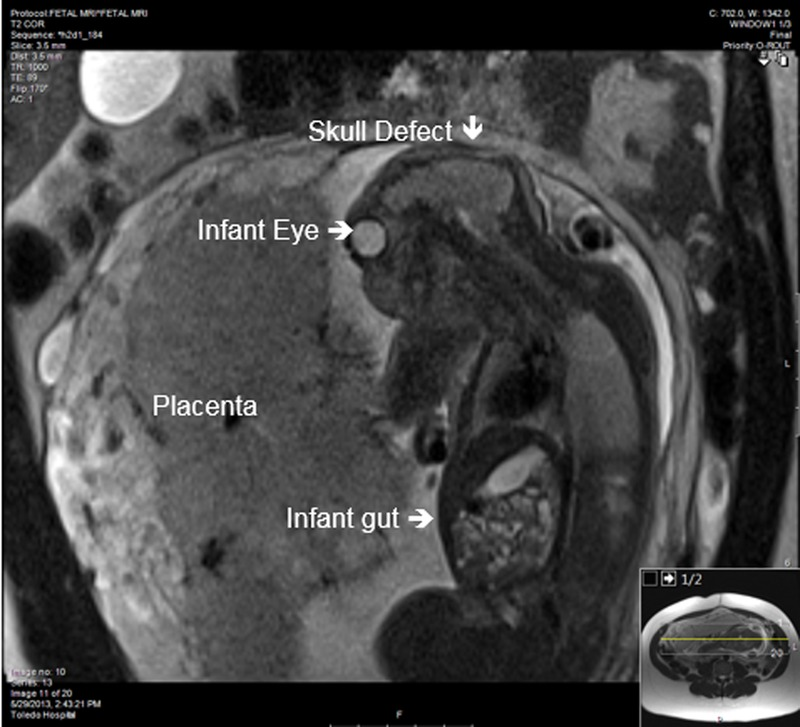
  Last semester, I was able to shadow a family medicine doctor, and I was presented with this rare case of a pregnancy that was anencephalic. Anencephalic is a congenital disorder, a birth defect that causes the baby to not develop a brain or skull (Cleveland Clinic, 2023). Studies have shown that this is such a rare case, 1 in 4,600 babies are diagnosed with anencephaly in the United States, however this is because this disorder affects the nervous system by not developing the neural tube that forms the brain and skull in the first month of pregnancy (CDC, 2023). The brain being essential to the body, and need for life as it pertains to functions like memory, body movement, and emotion. 

    The diagnosis for anencephaly is still unknown, but it is usually detected within the pregnancy though an ultrasound or blood test suggesting that there are levels of high alpha-fetoprotein, opioid use, and high heat exposure ( Cleveland Clinic, 2023). There are three different types of anencephaly: meroacrania, holoanencephaly, and acrania. According to Children’s Hospital, meroacrine is the milder form of this disorder and small area of the skull missing, acrania is the entire skull is missing and part of the brain is developed: while the holoprosencephaly is where the entire brain is missing (Children's Hospital Colorado, 2023). 


    Therefore, there seems to be a rare case for survival of babies with this congenital disorder, but there was one case that lived up to 28 months. This baby was a female infant born by C-section at 40 weeks and 2 days of gestation. As she reached the 28 months of life, she continuously got check ups, and it was noticed that the infant slept during the day and had an irregular heartbeat ( PubMed Central, 2016). This infant would smile occasionally and make cooing noises, but would obtain seizures as she got closer to the end of her life (National Library of Medicine, 2016). This was in result to not having a brain that allowed her to have emotions, form memories; but instead due to no structure in the brain, she obtained a lot of seizures.

    As this case was rare, it became information that defied the current research on babies with this birth defect, that gave us a glimpse of the physiological effects that anencephalic disorder brings to the medical field. There are no treatments for this congenital disorder, and it is recommended to have a support system that can help with a family member with this condition.

Work Cited:

Anencephaly. Children’s Hospital Colorado. (n.d.). https://www.childrenscolorado.org/conditions-and-advice/conditions-and-symptoms/conditions/anencephaly/

Centers for Disease Control and Prevention. (2023, June 28). Facts about anencephaly. Centers for Disease Control and Prevention. https://www.cdc.gov/ncbddd/birthdefects/anencephaly.html

Dickman, H., Fletke, K., & Redfern, R. E. (2016, October 31). Prolonged unassisted survival in an infant with anencephaly. BMJ case reports. https://www.ncbi.nlm.nih.gov/pmc/articles/PMC5093842/

professional, C. C. medical. (n.d.). What is anencephaly?. Cleveland Clinic. https://my.clevelandclinic.org/health/diseases/15032-anencephaly 

Caffeine: Healthy Or Hazardous?

     Caffeine is a common stimulant that is widely accepted in our society as being normal, it is able to stimulate the central nervous system within an individual to decrease fatigue. These caffeinated drinks that are widely produced promise increased energy, improved physical performance, and a higher mental function in adults. How about children? Is there something more to these drinks as they seem, as many call them, "too good to be true." This blog will focus on the effects that caffeine can have on both adults and children and if they are safe for the younger generation to consume. 

    For caffeine to be safe and effective in adults it must be used correctly. When it is not used correctly it is  seen to effect sleep patterns, increased anxiety, and can produce other negative health effects. Many adults will use caffeine regularly so that they may increase aerobic endurance and strength, improve reaction time, and delay fatigue, but how do they accomplish this? They use caffeine correctly, consuming about 3-6mg/kg. In some cases they may consume more than what is recommended, ingesting nearly 13 mg/kg. While this does increase ones performance, it will bring about negative effects once the caffeine finally vacates their system. This can lead to a large crash off of the caffeine, headaches, and trouble sleeping. Overall, in adults, it has been seen that caffeine can be very beneficial if it is used correctly.

    Caffeine in adolescents is an entirely different story. The American Association of Poison Control Centers reported more than 4600 calls and of these 4600 calls 2345 individuals needed treatment due to some type of caffeine toxicity or abuse. This is because of the serving size in many of these cans and bottles which caffeine can be found. As children are usually much smaller than adults, this results in a higher mg/kg than is recommended. This high concentration of caffeine can lead to anxiety and heart arrythmias in children. High doses of caffeine can also result in caffeine toxicity which can also lead to a list of negative impacts such as trouble breathing, vomiting, and chest pain. Caffeine has also showed to effect the developing neurologic and cardiovascular systems. This is a main concern for developing children as this can cause life long complications. Lastly, as children are very impressionable at their young ages, they are much more likely to become addicted to this substance as their bodies can easily become dependable on the energy it provides. If they do not fulfill their bodies desire, it can result in headache, fatigue, decreased alertness, drowsiness, difficulty concentrating, irritability, and depressed mood. The need for caffeine in children can be irradicated by simply providing them with a well balanced diet, as this will be able to provide the energy necessary for them to complete their daily tasks.

    Overall the consumption of caffeine by adults can be very beneficial to them if they consume the product in the correct measurements to their own body. But for adolescents it can have very devastating short term and long term effects. Children should not be drinking caffeine until they reach the correct age, in order to ensure full bodily development and to ensure they have the maturity necessary to ingest caffeine correctly.


Schneider, M. B., & Benjamin, H. J. (2011, June 1). Sports drinks and energy drinks for children and adolescents: Are they appropriate?. American Academy of Pediatrics. https://publications.aap.org/pediatrics/article/127/6/1182/30098/Sports-Drinks-and-Energy-Drinks-for-Children-and?autologincheck=redirected

What is Celiac Disease?

Celiac disease is a chronic digestive and autoimmune disorder that affects the small intestine and affects about 1% of the general population. It is caused by the ingestion of gluten, which is a major protein that is stored in wheat, rye, and barley. These proteins drive the immune response to celiac disease. Gastric and pancreatic enzymes are unable to break down the gluten proteins, which creates an autoimmune response. Celiac disease is caused by genetic factors and having a family history of celiac disease increases the risk of developing the disease by 10-15%. MHC class II genes are the genes responsible for the disease. 90% of patients with celiac disease express the gene, HLA-DQ2.5. The patients who are most at risk for developing the disease are homozygous with HLA-DQ2. Celiac disease can be hard to diagnose because it presents with any age and various symptoms. Because there are different clinical features at the time of diagnosis, celiac disease is broken into 3 forms: classical, non-classical, and subclinical. The classical form is most common in children under 5 years old, who present with “chronic diarrhea, poor appetite, weight loss, abdominal distension, muscle wasting, and mood changes” (Catassi et al., 2022). Non-classical is the most prevalent and consists of non-specific intestinal complaints, chronic fatigue, nutritional deficiencies, etc. Subclinical is diagnosed by screening programs or case finding in high risk groups, such as having a family history of celiac disease. 

Celiac Disease is also associated with other autoimmune diseases such as type 1 diabetes and Hashimoto’s thyroiditis. 4.5% of patients with type 1 diabetes will also have celiac disease. Testing for celiac disease involves using serological markers, HLA gene determination, and/or a small intestinal biopsy. The appearance of serum antibodies is a key factor in the diagnostic process. People who are screened for celiac and have a gluten-containing diet will show IgA class anti-TG2, which is the best screening test to start with. The most effective treatment for celiac disease is a major lifestyle change, which is to switch to a gluten-free diet. It is nutritionally safe and enables healing without major complications. Even though cutting out gluten from a diet is the most effective treatment, there is still ongoing research for new treatments such as nutritional support, pharmacological agents, and stem-cell transplantation. Further research is needed for new treatments and therapies that could hopefully improve the quality of life for patients with celiac disease. 


Catassi, C., Verdu, E. F., Bai, J. C., & Lionetti, E. (2022). Coeliac disease. The Lancet, 399(10344), 2413–2426. https://doi.org/10.1016/s0140-6736(22)00794-2 


THE PHYSIOLOGICAL EFFECTS OF PROLONGED WAR EXPOSURE

     We have all heard about the Israeli-Palestinian conflict in the news. Major events such as President Biden’s solidarity with Israel, and Israel bombing Palestinian hospitals (and threatening to bomb more, where injured individuals are already screaming in pain because these hospitals ran out of pain medication) have taken over our news feeds. It is no doubt that living in these areas under constant threat has a number of physiological effects on these individuals. Such as in soldiers, children, and mothers.


    Hamas and Israel have fought five wars since 2006 (The Economist Newspaper), meaning that individuals living in Israel and Palestinian territories have lived in a war zone for at least 17 years (there have been conflicts before, I encourage you to look into the history). In these war zone conditions, the sympathetic nervous system completely overrides the parasympathetic oversystem, preventing soldiers’ bodies from reaching and/ or maintaining homeostasis. Adrenaline continues to flow for hours on end, and in order to survive, soldiers naturally learn to block everything around them out. After living in these conditions for years, their bodies become used to living in “survival mode” and returning home without constant threats does not mean they automatically return to homeostasis. Their bodies can actually become even more off-balance. From the Iraq Afghanistan war, about 19% of soldiers return with PTSD but to avoid being perceived as “defective” only about half of these soldiers reach for help (Bruner et al., 2011). This war lasted from 2003-2011 (The Library of Congress) so it can be expected that these numbers will increase for the ongoing Isreali-Palestinian war.


    As mentioned earlier, hospitals with innocent women and children are being bombed and more are in constant threat of a bombing. Other than experiencing similar physiological effects of heightened awareness, women experience a decrease of fertility due to a reduction of sex hormones which can lead to anxiety, which can lead to depression (Bruner, et al., 2011). All the while, many of these women are left to care for their children alone. Children are hypersensitive to their surroundings and maternal influences, and in these conditions their HPA-axis is negatively affected. HPA-axis dysregulation affects the way their bodies respond to stress, and can lead to mental disorders in the future.


Of course the effects on soldiers, women, and children can overlap in different levels and there are plenty more physiological systems to take into account.


Bruner, V., & Woll, P. (2011). The battle within: Understanding the physiology of war-zone stress exposure. Social Work in Health Care, 50(1), 19–33. https://doi.org/10.1080/00981389.2010.513915 

The Economist Newspaper. (n.d.). Young Palestinians in Gaza cannot find work and cannot leave. The Economist. https://www.economist.com/middle-east-and-africa/2022/07/14/young-palestinians-in-gaza-cannot-find-work-and-cannot-leave 

Halevi, G., Djalovski, A., Kanat-Maymon, Y., Yirmiya, K., Zagoory-Sharon, O., Koren, L., & Feldman, R. (2017). The social transmission of risk: Maternal stress physiology, synchronous parenting, and well-being mediate the effects of war exposure on child psychopathology. Journal of Abnormal Psychology, 126(8), 1087–1103. https://doi.org/10.1037/abn0000307

Wars in Iraq (2003-2011) and Afghanistan (2001-2021)  :  serving: Our voices  :  veterans history project collection  :  digital collections  :  library of Congress. The Library of Congress. (n.d.). https://www.loc.gov/collections/veterans-history-project-collection/serving-our-voices/wars-in-iraq-and-afghanistan/


Nutrition and Exercise Physiology in Female Athletes

    Since the passing of the Title IX act in 1972, there has been a significant increase in involvement and engagement surrounding women's athletics. However, with all this development, there has been a consequential lack of research into female-specific areas of athletics, such as nutrition and exercise physiology. As we see in most areas of medicine, this lack of research is mostly due to the assumption that the female body is much more complicated to study due to hormonal implications. A female menstrual cycle can significantly impact what is going on physiologically in the rest of the body, and hormone cycles (specifically luteinizing hormone and follicle-stimulating hormone) can require longer periods of observation, to ensure all data is being collected. Due to all of these “complications” with research into the female body, there is little to no research into what a balanced nutrition looks like for a woman as well as how women should plan out and execute their exercise methods. However, there is some hope, as a 2021 review article from Holtzman and Ackerman investigated these exact issues and aimed to provide insight for female athletes. They looked heavily into how nutrition plays a significant role into the success of female athletes, and what their diet should look like based around their hormonal cycle. From this research, they suggested a few key things for women to consider when making a dietary plan. First, they state that women should pay close attention too their menstrual cycle in order to monitor for any deficiencies that may occur, then they suggested eating a wide range of foods in order to get the micronutrients that are necessary for the body to function, and the last main point they made in regards to diet was that women, and specifically female athletes, should be taking supplemental vitamins like iron and calcium in order to replenish what can be lost during menstruation. Overall, while this paper is beneficial in providing some insight into how the female body is different regarding athletics, nutrition, and exercise physiology, there is still a lot of research that needs to be done to help aid female athletes and the growth of the women’s game, as well as just the female population.


Holtzman, B., Ackerman, K.E. Recommendations and Nutritional Considerations for Female Athletes: Health and Performance. Sports Med 51 (Suppl 1), 43–57 (2021). https://doi.org/10.1007/s40279-021-01508-8

Caffeine in Children and Adolescents: Are energy drinks and sports drinks the same?

    Sports and energy drinks are a large and growing beverage industry that aim to market to children and adolescents for a variety of uses. Sports drinks suggest optimization of athletic performance and replacement of fluid and electrolyte loss. Whereas, energy drinks suggest energy boosts, decreased fatigue levels and enhanced concentration and mental alertness. The primary objective of this report is to help improve education on the differences between sports and energy drinks and how to eliminate inappropriate uses by children and young adults. 


Caffeine, the primary ingredient in energy drinks, has been shown to enhance physical performance in adults by increasing aerobic endurance and strength, improving reaction time, and delaying fatigue. However, these effects are extremely variable, dose dependent, and, most importantly, have not been studied in children and adolescents. Ergogenic effects have been reported with doses of 3 to 6 mg/kg, with some adult athletes reaching daily caffeine intakes of up to 13 mg/kg. Notable effects of caffeine intake are an increase in heart rate, blood pressure, speech rate, motor activity, attentiveness, gastric secretion, diuresis, and temperature. Caffeine can increase anxiety in those with anxiety disorders, play a role in triggering dangerous heart arrhythmias, and disturb regulated sleeping patterns.


The study noticed a heightened awareness of the risks of caffeine use, abuse, and even toxicity in children and adolescents. For example, in 2005, the American Association of Poison Control Centers reported more than 4600 calls regarding caffeine, with 2600 of these calls involving patients younger than 19 years old and 2345 patients requiring treatment.


Sports drinks have an important, specific role in the diet of young athletes who are engaged in prolonged vigorous sports activity—primarily to rehydrate and replenish carbohydrate, electrolytes, and water lost during exercise. However, the confusion about energy drinks versus sports drinks in children and adolescents can lead to unintentional ingestion of energy drinks. Using energy drinks instead of sports drinks for rehydration can result in ingestion of potentially large amounts of caffeine or other stimulant substances and the adverse effects previously described.


COMMITTEE ON NUTRITION AND THE COUNCIL ON SPORTS MEDICINE AND FITNESS. Adolescents: Are They Appropriate? Clinical Report – Sports Drinks and Energy Drinks for Children and Adolescents: Are They Appropriate? Pediatrics, 127 (6): 1182-1189, 2011. https://publications.aap.org/pediatrics/article/127/6/1182/30098/Sports-Drinks-and-Energy-Drinks-for-Children-and?autologincheck=redirected

Tuesday, October 17, 2023

Adjunctive LAT-Activating CAR T (ALA-CART) and Implications on Cancer Treatment


Cancer is more common than you think. It is the second leading cause of death, after heart disease in the U.S[1], and a leading cause of death globally.[2] In developed countries, 40-50% of people will get cancer in their lifetime[3], and of the 40-50% of people who get cancer, around 50% will be cured.[4] Traditional treatments for cancer include chemotherapy, surgery, and radiation.[5] Traditional treatments have several limitations, for instance, surgery and radiation can only remove or kill cancer cells in localized tumors but cannot eliminate tumor cells that have left the primary tumor site. Chemotherapy works by killing rapidly dividing cells, including tumor cells but also healthy cells that rapidly divide. The limitations of traditional therapies lead to treatment failures and relapses which cause the majority of cancer-related deaths. Therefore, there is a need for new therapies for cancer, including immunotherapy which utilizes the body’s T-cells.

Over the summer, I did research with the Kohler Lab on a type of cancer immunotherapy treatment called Chimeric Antigen Receptor (CAR) T-cell. It is a treatment where T-cells from a patient are genetically modified so they will target cancerous cells through the recognition of an antigen expressed on the surface of cancer cells by the Chimeric Antigen Receptor. CAR T-cells have had multiple successes in patients. Currently, there are six FDA-approved CAR T-cell therapies treatment. Four of those target CD19 antigens, and two of those target BCMA.[6] However, there are limitations in CAR T-cell therapies which lead to relapses. Those are short persistence and decreased sensitivity to low levels of antigens. Those limitations are predicted to stem from a weakening of LAT signaling, which is an important molecule in T-cell’s activation mechanism.

To combat these limitations, the Kohler Lab is researching on Adjunctive LAT-activating CAR T-cell (ALA-CART). ALA-CART places a LAT CAR next to a standard CAR to force the interaction between the LAT molecule and the CAR T-cell[7], instead of relying on endogenous LAT molecules. LAT molecule is an important molecule in T-cell's activation mechanism. So far, ALA-CART has shown promising results, which enhances antigen sensitivity and persistence of CAR T-cells.[8],[9]


[1] Centers for Disease Control and Prevention. An Update on Cancer Deaths in the United States. Atlanta, GA: US Department of Health and Human Services, Centers for Disease Control and Prevention, Division of Cancer Prevention and Control; 2022. https://www.cdc.gov/cancer/dcpc/research/update-on-cancer-deaths/index.htm

[2] World Health Organization. Cancer. Feb 3 2022. https://www.who.int/news-room/fact-sheets/detail/cancer

[3] American Cancer Society. Cancer Facts & Figures 2022. Atlanta: American Cancer Society; 2022.

[4] American Cancer Society. Cancer Facts & Figures 2022. Atlanta: American Cancer Society; 2022.

[5] MedlinePlus [Internet]. Gersten, Todd (MD): National Library of Medicine (US); [updated Oct 28 2021; cited 2023 Jul 16]. Available from: https://medlineplus.gov/ency/patientinstructions/000901.htm#:~:text=If%20you%20have%20cancer%2C%20your,%2C%20hormonal%20therapy%2C%20and%20others.

[6] National Cancer Institute: “CAR T Cells-Engineering Patients’ Immune Cells to Treat Their Cancers”. Updated March 10 2022 https://www.cancer.gov/about-cancer/treatment/research/car-t-cells


[7] University of Colorado Cancer Center: Eric Kohler, MD, PhD, Receives Award to Improve CAR T-cell Therapy”. Feb 24, 2023. https://news.cuanschutz.edu/cancer-center/eric-kohler-ash-scholar-award


[8] Danis, Catherine, et al. 2023. https://doi.org/10.1158/1538-7445.AM2023-3190

[9] Danis, Catherine, et al. 2022. https://doi.org/10.1158/1538-7445.AM2022-3607


Friday, October 13, 2023

Deadly Duo

As most know, the leading cause of death in developed and developing countries is coronary heart disease (CHD). CHD occurs when plaque is formed within the coronary arteries and therefore has a difficult time distributing blood and oxygen to the cardiac muscle tissue. Over recent years there has been seen an increased correlation between atrial fibrillation (AF) and the cause of CHD and vice versa due to their common risk factors. CHD can cause AF due to reentry formation, focal ectopic activity, and neural remodeling, all of which are common cardiac arrhythmia origins. To me, the most interesting cause here is the neural remodeling in which the autonomic nervous system change’s function due to CHD and therefore causes AF. On the other hand, AF can cause CHD via atherosclerosis, mismatched blood supply and oxygen consumption, and thrombosis by changing the heart’s ability to affectively beat. Interestingly enough, atherosclerosis from AF has been hypothesized to cause CHD because of the increased von Williebrand factor in these patients, making their blood vessels weaker and narrowing the blood supply potential. In this journal entry it was found that patients that already had AF developed CHD 34% of the time. With this comorbidity much worse outcomes occur, such as myocardial infarctions and even death.  Although there is a clear and proven correlation between CHD and AF, the exact mechanisms for their causation are still unknown. What research needs to be done to break up this “vicious cycle”? Knowing that one disease causes the other, if we can prevent either from their initial onset, then the comorbidity will never occur. 

As someone who has a family lineage of heart disease it is important for me to understand this dual relationship and watch out for the common risk factors more than the average person. Some of these risk factors include: hypertension, diabetes, obesity, and sleep apnea. I can combat these factors by maintaining a healthy lifestyle and focusing on heart healthy foods and exercise. Knowing that I am higher risk to develop heart disease and therefore AF, what further research can be done in terms of preventative care for not only me but others who suffer from similar genetic backgrounds? 


Source: Liang, F., & Wang, Y. (2021). Coronary Heart Disease and Atrial Fibrilation: A Vicious Cycle. In American Journal of Physiology-Heart and Circulatory Physiology (Vol, 320, Issue 1, pp. H1-H12). American Physiological Society. https://doi.org/10.1152/ajpheart.00702.2020

What is Diabetic Retinopathy?

Working as an optometric technician for the past year, I was able to familiarize myself with ocular diseases. There were patients that would come in for eye exams specifically to check on their ocular health because they were diabetic. Diabetes, a metabolic disorder that causes an imbalance in blood glucose levels, can be accompanied by diabetic retinopathy (DR), which can lead to blindness (Sapra & Bhandari, 2022; Jenkins et al., 2015). Out of the 463 million people that have diabetes, 126 million have diabetic retinopathy (Saeedi et al. 2019; Jenkins et al. 2015).
 
Diabetic retinopathy occurs when there is a problem with one’s vasoregulation, meaning blood flow in the vessels becomes dysfunctional (Chen et al., 2022). These vasoregulation problems are caused by the increase in glucose in the body, which affects the blood vessels in the retina, the inner layer of the eye where light is converted to images (Mahabadi & Khalili et al., 2023). On the retina is the macula, where an individual’s most sensitive and accurate vision occurs; thus, if an individual has improper vascular flow, fluid can build up and push on the retina (Chen et al., 2022). These physiological disruptions can lead to diminished vision or vision loss.
 
Diabetic retinopathy can be manageable if diagnosed early on. Diagnosing DR can include taking ultra wide fundus photos of the retina, allowing optometrists or ophthalmologists to easily see the majority of the retina, and using Optical Coherence Tomography, which images a cross section of the retina (Park & Roh, 2016). The early stage of DR is called non-proliferative diabetic retinopathy and is characterized by leaking of blood vessels, causing microaneurysms and possible swelling of the macula (Wang & Lo, 2018). The later stage of DR is called proliferative diabetic retinopathy and can worsen vision problems due to hemorrhaging in the eye and retinal detachment (Wang & Lo, 2018).
 
After diagnosing the diabetic retinopathy, discussing treatment methods is critical as to not further one’s vision loss. One of the most common treatments is the use of laser photocoagulation, which prevents further vision loss but does not restore it (Wang & Lo, 2018). Less frequently used are intravitreal steroids, and, in advanced cases of DR, surgery is performed (Wang & Lo, 2018). It is important for diabetics to visit their optometrist or ophthalmologist to monitor their ocular health.

References

Chen, Y., Coorey, N. J., Zhang, M., Zeng, S., Madigan, M. C., Zhang, X., Gillies, M. C., Zhu, L., & Zhang, T. (2022). Metabolism Dysregulation in Retinal Diseases and Related Therapies. Antioxidants (Basel, Switzerland), 11(5), 942. https://doi.org/10.3390/antiox11050942

Jenkins, A. J., Joglekar, M. V., Hardikar, A. A., Keech, A. C., O'Neal, D. N., & Januszewski, A. S. (2015). Biomarkers in Diabetic Retinopathy. The review of diabetic studies : RDS, 12(1-2), 159–195. https://doi.org/10.1900/RDS.2015.12.159

Mahabadi N, Al Khalili Y. Neuroanatomy, Retina. [Updated 2023 Aug 8]. In: StatPearls [Internet]. Treasure Island (FL): StatPearls Publishing; 2023 Jan-. Available from: https://www.ncbi.nlm.nih.gov/books/NBK545310/

Park, Y. G., & Roh, Y.-J. (2016). New diagnostic and therapeutic approaches for preventing the progression of diabetic retinopathy. Journal of Diabetes Research, 2016, 1–9. https://doi.org/10.1155/2016/1753584 

Saeedi, P., Petersohn, I., Salpea, P., Malanda, B., Karuranga, S., Unwin, N., Colagiuri, S., Guariguata, L., Motala, A. A., Ogurtsova, K., Shaw, J. E., Bright, D., Williams, R., & IDF Diabetes Atlas Committee (2019). Global and regional diabetes prevalence estimates for 2019 and projections for 2030 and 2045: Results from the International Diabetes Federation Diabetes Atlas, 9th edition. Diabetes research and clinical practice, 157, 107843. https://doi.org/10.1016/j.diabres.2019.107843

Sapra A, & Bhandari P. Diabetes Mellitus. [Updated 2022 Jun 26]. In: StatPearls [Internet]. Treasure Island (FL): StatPearls Publishing; 2023 Jan-. Available from: https://www.ncbi.nlm.nih.gov/books/NBK551501/ 

Wang, W., & Lo, A. C. Y. (2018). Diabetic Retinopathy: Pathophysiology and Treatments. International journal of molecular sciences, 19(6), 1816. https://doi.org/10.3390/ijms19061816